Medical imaging technology plays a major role in how doctors understand what is happening inside your body without making a single cut.
When I first learned about it, I was surprised by how much these tools can reveal with just a quick scan or picture.
In this post, I’ll walk you through what medical imaging is, why it matters, and how it helps doctors make better decisions for you.
You’ll see the main types of imaging, how each one works, and the key benefits that make them so useful. I’ll keep everything simple, clear, and easy to follow, so you never feel lost in technical terms.
By the end, you’ll have a solid picture of how these methods support early detection, safer care, and more accurate treatment.
My guide gives you the basics you need to understand them with confidence.
What Does Medical Imaging Mean?
Medical imaging means using special machines to create pictures of the inside of your body without surgery.
These pictures help doctors understand what’s going on when they can’t see the problem from the outside. With X-rays, CT scans, and MRIs, doctors can see bones, organs, and tissues safely and clearly.
Medical imaging is important because it shows details that exams alone can’t reveal.
It helps find injuries, track illnesses, and guide treatment with more accuracy.
This technology gives doctors the information they need to choose the right steps for your care. Instead of guessing, they can see what’s happening and act quickly.
Medical imaging also reduces the need for risky procedures, since many answers come from a simple scan.
It’s a direct, practical method that supports better and safer healthcare.
How Medical Imaging Works?
Medical imaging uses different machines to create clear pictures of what’s happening inside your body. Each tool works in its own simple way to help doctors see bones, organs, and tissues without surgery.
- X-rays use light waves to pass through your body and capture quick pictures of bones and dense tissues.
- CT scans take many X-ray images from different angles and combine them to form a detailed, 3D view.
- MRIs use strong magnets and radio waves to make sharp images of organs, muscles, and soft tissues.
- Ultrasound uses sound waves that bounce off tissues to create real-time images, often used for pregnancy and organ checks.
- Nuclear imaging uses small amounts of tracers to show how organs and tissues are working, not just how they look.
Main Types of Medical Imaging
These imaging methods help doctors see inside your body in different ways, depending on what needs to be checked. Each type offers unique details that support diagnosis, treatment, and follow-up care.
1. X-Ray
X-ray imaging uses a small amount of radiation to create clear pictures of bones and dense tissues.
It helps doctors find fractures, infections, or lung issues quickly. Because the process is fast, X-rays are often the first test used when someone has an injury or sudden pain.
They show strong detail in hard structures but less detail in soft tissues.
Even with limits, X-rays remain one of the most common and useful imaging tools in healthcare.
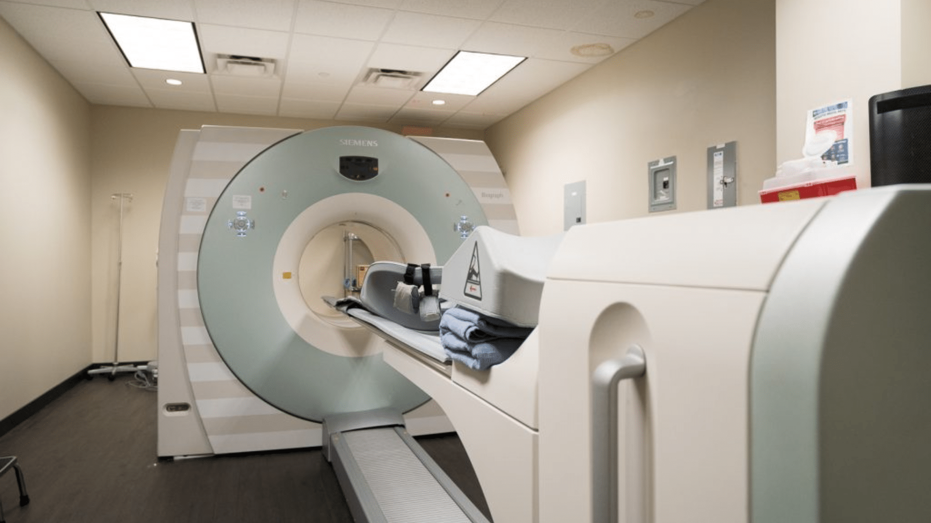
2. CT (X-Ray Computed Tomography)
CT scans take many X-ray images from different angles and combine them into one detailed, cross-sectional view.
This helps doctors see bones, organs, and tissues with far more clarity than a regular X-ray. CT is useful for spotting internal injuries, tumors, infections, and bleeding.
The images load quickly, making CT a helpful tool during emergencies.
Though CT uses more radiation than X-rays, it provides a deeper, more complete picture of what’s happening inside the body.
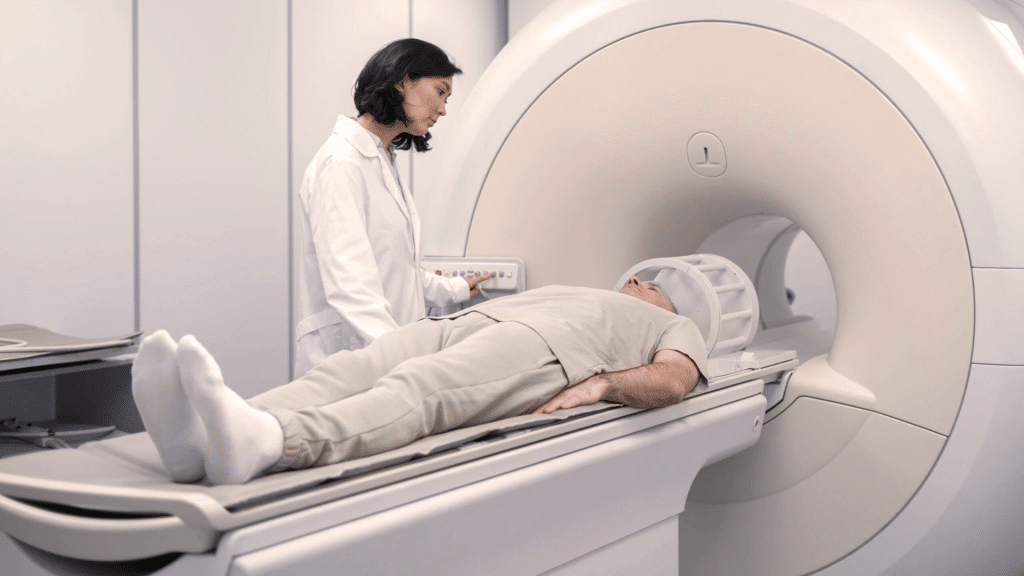
3. MRI (Magnetic Resonance Imaging)
MRI uses strong magnets and radio waves to create detailed pictures of soft tissues like the brain, muscles, nerves, and organs.
It does this without using any radiation, which makes it safer for repeated scans.
MRI is especially helpful for finding brain issues, joint injuries, and spinal problems. The images are sharp and full of detail, giving doctors precise information.
Although MRI takes longer and can be noisy, it offers a level of clarity that many other tests cannot match.
4. PET (Positron Emission Tomography)
PET imaging shows how organs and tissues are working by using a small tracer that highlights activity inside the body.
Instead of only showing structure, PET reveals function, which helps doctors spot cancer, track treatment progress, and study brain and heart activity.
It gives a clear picture of how cells behave, not just how they look.
PET is often paired with CT to combine functional and structural details, allowing doctors to make better treatment plans with more accurate information.
5. SPECT (Single-Photon Emission Computed Tomography)
SPECT imaging uses a radioactive tracer to show blood flow and activity in organs and tissues.
It provides 3D images that help doctors understand how well the heart, brain, and bones are working. SPECT is commonly used to detect heart disease, track seizures, and examine bone conditions.
While it shares some features with PET, SPECT is more widely available and often more affordable.
It offers valuable insight into the body’s function, giving doctors information they can’t get from structural scans alone.
6. fMRI (Functional Magnetic Resonance Imaging)
fMRI measures brain activity by tracking changes in blood flow. When a part of the brain works harder, it receives more oxygen, and fMRI captures this shift.
The images help doctors and researchers understand how different brain areas support movement, memory, speech, and emotions.
fMRI is also useful for planning surgeries by showing which areas must be protected.
It provides real-time insight into how the brain functions, offering information beyond what a standard MRI can show.
7. DTI (Diffusion Tensor Imaging)
DTI is a special form of MRI that maps how water moves along nerve pathways.
By studying this movement, DTI shows the structure and health of white matter in the brain. It helps doctors see whether nerve fibers are damaged from injury, stroke, or disease.
DTI is especially useful for planning brain surgery and understanding conditions that affect communication between brain regions.
It gives a deeper look at wiring inside the brain, offering details that a regular MRI cannot provide.
8. 3D (Three-Dimensional Imaging)
3D imaging creates layered, lifelike pictures of organs and tissues, giving doctors views from many angles.
It is widely used in CT, MRI, and ultrasound to make diagnoses easier and more accurate. Surgeons often rely on 3D images to plan procedures with greater precision.
The depth and detail help doctors understand complex structures, such as tumors or fractures, in a clearer way.
3D imaging improves accuracy, reduces guesswork, and supports safer medical decisions for many types of care.
9. AI (Artificial Intelligence)
AI supports medical imaging by helping computers analyze scans quickly and accurately.
It can spot patterns, flag concerns, measure changes, and assist doctors in making decisions. AI reduces the time needed to read images and helps catch details that may be hard to see.
It also improves consistency across reports. While AI does not replace doctors, it strengthens their work by offering fast, data-driven support.
This leads to better detection, fewer errors, and more efficient healthcare.
10. ML (Machine Learning)
Machine learning is a type of AI that learns from data to improve image analysis over time.
In medical imaging, ML algorithms study thousands of examples to recognize patterns linked to diseases. This helps doctors detect problems earlier and with more accuracy.
ML is used in CT, MRI, ultrasound, and other tools to classify tissues, measure growth, and compare changes across scans.
Its ability to learn and improve makes it a powerful partner in modern imaging.
11. XAI (Explainable Artificial Intelligence)
XAI focuses on making AI decisions clear and easy to understand.
In medical imaging, this means doctors can see why an algorithm flagged a spot, measured a change, or suggested a diagnosis.
XAI builds trust by showing the reasoning behind each result. It helps doctors confirm whether the AI’s findings make sense and prevents confusion or hidden errors.
By offering transparency, XAI supports safer and more reliable use of AI tools in patient care.
12. CLAIM (Checklist for Artificial Intelligence in Medical Imaging)
CLAIM is a checklist that guides how AI research in medical imaging should be reported.
It ensures studies are clear, complete, and trustworthy. CLAIM helps researchers describe how their AI tool was built, tested, evaluated, and validated.
This makes it easier for doctors, hospitals, and other researchers to judge reliability and compare results.
With CLAIM, AI studies follow consistent standards, improving safety and transparency. It plays an important role in developing dependable AI tools for imaging.
When Medical Imaging is Used?
Medical imaging is used when doctors need to understand what’s happening inside your body without surgery.
It helps diagnose injuries like broken bones, joint problems, or internal bleeding.
Imaging also supports tracking long-term conditions, such as heart disease, cancer, or lung issues, by showing changes over time.
Doctors often use it to guide treatment, making sure procedures like biopsies or injections are placed in the right spot. It’s useful in emergency care when quick answers are needed.
Imaging also helps monitor recovery after surgery or illness, giving a clear view of how well your body is healing.
Each scan provides information that doctors can’t get from a physical exam alone.
Key Benefits of Medical Imaging
Medical imaging offers many advantages that help doctors understand health problems quickly and safely. These benefits support better decisions, early care, and more accurate treatment for you.
1. Early Detection
Medical imaging helps doctors find problems early, sometimes before symptoms appear.
This gives you a better chance of getting the right care at the right time. Early detection can reveal issues like small tumors, infections, or hidden injuries that might be missed in a physical exam.
With clearer pictures, doctors can act sooner and prevent conditions from getting worse.
This early look inside your body often leads to safer treatment and better results for many health concerns.
2. Accurate Diagnosis
Accurate diagnosis is one of the biggest strengths of medical imaging. These tools show clear details of bones, organs, and tissues, helping doctors identify the exact issue instead of guessing.
With precise images, they can spot fractures, tumors, infections, or blocked blood vessels.
This reduces mistakes and supports confident decision-making.
When doctors understand the problem fully, they can choose the treatment that fits their needs. Imaging also saves time by ruling out conditions that don’t match the results.
3. Less Invasive Care
Medical imaging reduces the need for invasive procedures by giving doctors the information they need without surgery.
Instead of exploring inside the body, doctors can use images to see what’s wrong and decide the next steps.
This lowers the risk of pain, infections, and complications. Imaging also guides procedures like biopsies, making them safer and more accurate.
When fewer invasive tests are needed, recovery time is shorter, and overall care becomes more comfortable and efficient for you.
4. Better Treatment Planning
Clear imaging helps doctors create treatment plans that match your specific condition.
These images show the size, location, and severity of a problem, giving doctors a full picture before acting. Good planning leads to safer surgeries, targeted therapies, and better long-term results.
It also helps doctors track how well your treatment is working and make adjustments when needed.
By using detailed images, doctors avoid guesswork and make decisions based on real, visible information from inside your body.
How Doctors Choose the Right Imaging Test?

Doctors choose the right imaging test by looking at your symptoms, medical history, and how quickly answers are needed.
They start with simple options when possible, such as X-rays for bone injuries or chest concerns. If more detail is required, they may order a CT scan to check for internal bleeding, infections, or organ problems.
MRI is chosen when doctors need clear views of soft tissues, the brain, or joints.
Your health background also matters, especially if you have allergies, kidney issues, or metal implants.
Urgency plays a big role, too, since some tests provide results faster than others. Each choice is made to get the most helpful information with the least risk.
Safety and Risks to Know
Medical imaging is generally safe, but each test has its own guidelines to protect you. Most scans have low risks when used correctly and for the right reasons.
- Radiation exposure is usually low, especially with X-rays and CT scans, and doctors only order these tests when needed.
- MRI uses no radiation, but you may need screening for metal implants or devices before the scan.
- Ultrasound is very safe, using sound waves instead of radiation.
- Preparation steps vary, such as fasting, removing metal items, or wearing loose clothing.
- Contrast dyes may be used, and your doctor will check for allergies or kidney issues first.
- Most imaging tests are considered low risk because they offer important information without surgery.
- Doctors weigh benefits and risks, choosing the safest test for your condition.
Advances in Medical Imaging Today
Advances in medical imaging today focus on making tests faster, clearer, and more comfortable for you. Many systems now use AI-assisted tools that help doctors read scans with better accuracy and fewer delays.
These tools can highlight areas that need attention, giving doctors stronger support when making decisions.
New machines also create sharper images, allowing doctors to see small details in organs, bones, and tissues.
Scans are quicker, so you spend less time in the machine. Updated designs make the process quieter and less cramped, which helps reduce stress.
Safety has improved as well, with lower radiation levels in tests that rely on X-rays or CT imaging. These improvements give doctors clearer information while keeping the experience safer and easier for you.
Overall, modern imaging provides better care with less discomfort and greater confidence in the results.
Conclusion
Understanding the different types of medical imaging technology helps you see how doctors find clear answers without relying on surgery.
In this post, you learned how each imaging tool works and why it matters for diagnosis and treatment.
These scans support early detection, safer care, and better treatment planning, which can make a big difference in your overall health.
When you know what each test does, you can feel more informed and less unsure if your doctor recommends one.
This knowledge also helps you understand the choices doctors make and why certain scans fit specific concerns. With this knowledge, you can be more confident and involved in your own health decisions.